The door closes with the gentle thump of a well-oiled hinge, and a familiar pattern settles in. Blood pressure cuff inflates, ultrasound warms on the cart, compression stockings wait in their sterile package. This is the rhythm of a modern vein practice, where a vein treatment specialist spends most days helping patients walk out lighter, with less pain and better legs than they brought in.

When people picture a vein and vascular doctor, they often imagine a surgeon in an operating theater. The real scene is quieter, more precise, and almost always performed under local anesthesia. The tools are slender catheters, a handheld ultrasound probe, and numbing solution, not scalpels and general anesthesia. The patient is awake, talking, sometimes laughing, and home within an hour.
Who does this work and how they train
Interventional vein care sits at the intersection of vascular medicine, interventional radiology, and surgery. Many of us trained first as vascular surgeons, interventional radiologists, or internists with additional specialization in venous disease. Some pursue board certification through the American Board of Venous and Lymphatic Medicine, while others carry boards in vascular surgery or interventional radiology and devote their practice to venous interventions. Titles vary, but whether you say venous specialist doctor, vascular vein specialist, or comprehensive vein doctor, the skill set centers on diagnosis with duplex ultrasound, minimally invasive procedures, and longitudinal management of chronic venous disorders.
Patients use a wide range of terms to find us. They search for a leg vein specialist when aching, bulging veins limit activity, a spider vein specialist when ankle webs spread, or a doctor for venous ulcers when a nonhealing sore keeps draining. Referrals come from primary care, wound clinics, podiatrists, and dermatologists. What unites the work is a focus on venous circulation and its failures: reflux, obstruction, inflammation, and clot.
What actually brings patients in
Symptoms tend to sound ordinary until you have lived them. Heaviness that builds across the day. A tugging pain behind the knee in the grocery aisle. Ankle swelling that leaves sock imprints like clay. Night cramps and restless legs that ruin sleep. The skin above the ankle that darkens to a coffee stain, then hardens and breaks down. Others notice only the visible veins, a rope of blue across the thigh or delicate red threads near the ankle.
We evaluate all of this under the umbrella of chronic venous insufficiency. As a vein disease expert, I look for three big patterns. First, venous reflux, the backward flow from faulty valves in superficial and perforator veins. Second, obstruction in deep veins, often from old clots or compression in the pelvis, such as May-Thurner anatomy. Third, inflammation in superficial veins, phlebitis, that flares and settles but leaves a tender cord. What looks cosmetic often has a hemodynamic driver. Treating only the surface without addressing reflux is like painting a damp wall.
The consult and mapping ultrasound
The first appointment is not a sales pitch. It is a clinical evaluation. We review symptoms, exercise habits, pregnancies, prior clots or leg trauma, and medications that thin blood. I examine the legs standing and supine, look for ankle skin changes, and palpate pulses. Then we study the system with ultrasound while the patient stands on a platform.
A trained vein ultrasound specialist walks the probe from groin to ankle. We measure vein diameters at defined points, test valves with compression and Valsalva, and document reflux duration in milliseconds. We map tributaries, perforator veins that connect surface to deep, and any tortuosity that limits catheter passage. The deep system is assessed for patency, residual thrombus, or scarring. For pelvic symptoms like heaviness with menses or vulvar varices, we sometimes extend imaging to the iliac veins with cross sectional studies or intravascular ultrasound during a later procedure.
Good mapping drives good outcomes. It tells a venous insufficiency specialist which segment needs closure, where to enter safely, and whether to add phlebectomy or foam. If the deep system is obstructed, closing a superficial pathway can worsen swelling. This is where judgment, experience, and a full vascular view count. A narrow focus on one vein misses the forest.
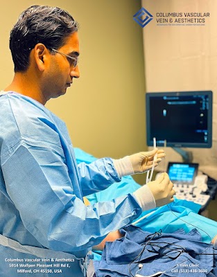
Inside the procedure room
Procedure day is engineered for calm. Patients do not fast unless sedation is planned, which is rare. We mark the leg with a skin pen, using the ultrasound map as a blueprint. The nurse starts a small IV if we anticipate sedation or fluids, then positions the patient on the table, often slight reverse Trendelenburg to help veins fill.
The room is small by design. Within an arm’s reach, I have the ultrasound unit, a cart with sterile packs, tumescent anesthesia, and the chosen catheter. The atmosphere matters. Music at low volume, warm blankets, and clear, simple steps reduce anxiety better than any pill.
Here is how a typical thermal ablation for a refluxing great saphenous vein proceeds:
- Site prep and drape. We scrub with chlorhexidine, lay sterile drapes, and expose only what we need. Access. Under ultrasound, I puncture the target vein with a micropuncture needle. The entry is usually below the knee for the great saphenous. We pass a soft guidewire, then a small sheath, much like placing an IV but with millimeter precision. Catheter positioning. The ablation catheter advances up the vein to a point 1.5 to 2.5 cm from the saphenofemoral junction. This margin protects the deep system. I confirm position in multiple planes. Tumescent anesthesia. Using a long infiltration needle, we bathe the vein along its length in a dilute lidocaine solution with epinephrine and bicarbonate. The tumescent fluid numbs tissue, compresses the vein around the catheter, and insulates skin and nerves from heat. This step takes the longest, and patient comfort hinges on steady hands and gentle technique. Energy delivery. We treat in small pulls, either by segmental radiofrequency or continuous pullback laser, depending on the system. The vein collapses and seals. The patient might feel warmth or a deep buzz, not pain. Adjuncts. If bulging tributaries remain, we often remove them through pinhole nicks, ambulatory phlebectomy. These micro-incisions do not need stitches and heal to faint dots. Sometimes we inject foam sclerosant under ultrasound into feeder branches. Compression and walk. We place a stocking or layered wraps and escort the patient to the hallway. A few minutes of walking stimulates calf pump and reduces clot risk.
The whole sequence takes 30 to 60 minutes. Patients leave upright, usually without narcotics. They can drive if we have not given sedatives. The plan that looked like alphabet soup on the first visit, EVLA, RFA, UGFS, now lands as a set of physical sensations that make sense.
Not one tool, but a toolkit
A vein management doctor does not sell a single box. We match methods to anatomy, symptoms, and patient goals.
- Thermal ablation, radiofrequency or endovenous laser, works well for long, straight refluxing trunks like the great or small saphenous veins. It has strong evidence for durable closure and symptom relief. Nonthermal, nonsclerosant closure, such as medical adhesive, avoids tumescent anesthesia and can help when a patient cannot tolerate multiple needle sticks. There are trade offs. You avoid heat near nerves, but you accept a foreign material and a different post procedure feel. Adhesive is useful in tortuous segments where getting a catheter through would be hard. Mechanochemical ablation combines a rotating wire with liquid sclerosant. It excels in veins close to the skin where heat could cause burns, and in patients who dislike tumescent volumes. It is less effective in very large diameter segments. Ultrasound guided foam sclerotherapy, whether physician compounded or with premixed canisters, can close smaller refluxing branches and perforators and treat residual veins after truncal closure. It is an art to move the foam by leg elevation, compression, and strategic injections so it fills the target without escaping. Ambulatory phlebectomy removes bulging tributaries through tiny nicks. It provides immediate debulking and cosmetic improvement. It does not treat axial reflux on its own.
If deep venous obstruction drives swelling, the conversation shifts. A peripheral vascular doctor might stent a compressed iliac vein with intravascular ultrasound guidance, especially in post thrombotic limbs. Treating superficial reflux alone would underdeliver.
Safety, risk, and how we keep complications rare
No procedure is risk free. We earn our keep by anticipating problems and structuring the day to avoid them. For thermal ablation, the main risks include skin burns, nerve irritation, bruising, superficial thrombophlebitis, and propagation of clot into the deep system. The last has a name, endothermal heat induced thrombosis, and we screen for it at follow up ultrasound. With proper technique, the rate of deep vein thrombosis after ablation is low, generally a few per thousand. Nerve symptoms are usually transient, affecting the saphenous nerve near the knee or sural nerve near the ankle after small saphenous work. Meticulous tumescent infiltration and careful catheter positioning make a difference.
Sclerotherapy carries risks of matting, a blush of fine vessels near the injection site, hyperpigmentation, transient visual aura in sensitive individuals, and rare allergy. Using the right concentration, slow injections, and immediate post treatment walking reduces these events. Adhesive closure can cause a focal inflammatory response that looks alarming but settles with time and NSAIDs.
I pay special attention to several scenarios:
- Anticoagulation. A circulation specialist doctor will not necessarily stop blood thinners. Thermal ablation and phlebectomy can proceed with careful hemostasis, but heavy phlebectomy under full anticoagulation is a recipe for bruising. We balance clot risk and bleeding case by case, involving the prescribing physician. Obesity. Access and catheter passage become harder with deeper veins, and tumescent volumes increase. Extra hands and longer needles help. Compression after the procedure is trickier to fit but essential. Pregnancy and postpartum. We avoid elective procedures during pregnancy and defer until after breastfeeding when hormones settle. For acute superficial thrombophlebitis in pregnancy, we lean on compression, ambulation, and targeted guidance rather than intervention. Active ulceration. A doctor for venous ulcers treats the underlying reflux early. Thermal ablation plus compression speeds healing compared to dressings alone. Wound care continues in parallel. Prior deep venous thrombosis. A vein and artery doctor looks for synechiae and scarring that redirect flow. Superficial closure can still help, but only if the deep pathway is open enough to carry increased load.
Everything runs smoother when the same vein diagnostics doctor who read the initial ultrasound is in the room guiding the catheter. Real time imaging is our fluoroscopy.
What patients do before and after
Preparation is minimal. I ask patients to hydrate well, bring their compression stockings, and wear shorts. If anxious, they can take a mild anxiolytic with a ride home. Shaving is not necessary and can irritate skin.
Here is the short checklist we hand out on the first procedure day:
- Bring thigh high 20 to 30 mm Hg compression stockings, unless we provided a different plan. Eat a light meal and hydrate, unless we discussed sedation. Avoid heavy lotions on the leg, they make draping harder. Arrange a ride if taking sedatives. Take routine medications unless instructed otherwise.
After the procedure, walk. The old advice of bedrest after vein work belongs to an era of vein stripping. We want calf pumps firing and blood moving. Wear compression during the day for a week or two. Sleep without it unless swelling flares at night. Avoid long, motionless car rides or flights for several days. Gentle exercise is fine the next day, but hold off on hot yoga, saunas, and heavy squats for a week. Pain is generally a deep ache that peaks at day 3 to 5 and responds to NSAIDs and walking.
We schedule a follow up ultrasound within a week to confirm closure and check for EHIT, then another visit at 4 to 6 weeks to assess symptom change and plan any staged treatments. Cosmetic injections, if needed, come last.
Cases that stay with you
A contractor in his fifties came to me after his third course of antibiotics for a leg wound. He wore elastic sleeves from the pharmacy that rolled down and cut into his calf. Ultrasound showed Columbus Vascular Vein & Aesthetics vein doctor OH a 7 mm great saphenous vein with 1.2 seconds of reflux and a perforator feeding the ulcer bed. We closed the trunk with radiofrequency, ligated the culprit perforator with a single needle puncture, and removed several bulbous tributaries. He called two weeks later not to talk about pain, but to say he could kneel again to set tile, the ache had lifted, and the wound had dried to a scab.
A young mother, otherwise healthy, could not bear how her legs felt by 5 p.m. She had visible varicosities and diffuse spider veins around the ankles. The duplex map showed bilateral reflux with competent deep systems. We performed endovenous laser ablation of both great saphenous veins two weeks apart, then staged phlebectomy. Her ankle webs received low concentration sclerotherapy months later. She did not care about the hemodynamics, she cared about bedtime with her toddler without throbbing legs. When you match the plan to what matters to the person, the technical choices fade into the background.

I have also turned patients away for intervention. A runner with prominent surface veins but no reflux and no symptoms beyond visibility needed a cosmetic spider vein specialist, not truncal ablation. Another patient with severe leg swelling had both reflux and pelvic outflow obstruction from old clot. A quick superficial closure would have failed. We coordinated with a colleague, a vascular medicine doctor with deep venous expertise, to treat the iliac segment first.
Outcomes you can measure
We track patient reported scores and objective findings. The Venous Clinical Severity Score falls as edema softens and skin improves. Ulcers heal faster when reflux is treated. Quality of life scales shift by several points after truncal ablation and phlebectomy. Durable vein closure rates for radiofrequency and laser ablation exceed 90 percent at one year in most published series. Sclerotherapy outcomes depend more on vein size and concentration, with higher retreatment rates for larger segments.
That does not mean every leg looks perfect. Some bruising lingers for weeks. Pigmentation after sclerotherapy can take months to lighten. Rarely, we reintervene for recanalized segments or neovascularity. A chronic vein doctor lives with the fact that venous disease is chronic. We manage, monitor, and adjust.
What insurance covers and when
Medical necessity is not a slogan. A vein care provider documents it. Most insurers cover truncal ablation when patients have proven reflux on duplex, symptoms like pain or edema, and a trial of compression that did not resolve symptoms. Purely cosmetic spider veins are usually self-pay. We explain the difference between a doctor who treats varicose veins for symptom relief and a cosmetic vein specialist doctor who focuses on appearance. The lines blur at times, but honest documentation removes the guesswork.
How to choose a vein professional
Titles and advertising can confuse. Some clinics emphasize lasers and aesthetics, others look like interventional radiology suites. Training, volume, and philosophy show in the first visit. When friends ask how to choose, I offer a simple framework.
- Verify credentials. Look for a licensed vein doctor with board certification in vascular surgery, interventional radiology, or venous and lymphatic medicine. Ask who performs and interprets the ultrasound. A vein imaging specialist on site who collaborates with the operator improves results. Expect a full leg assessment. A certified vein specialist should examine you standing, map reflux, and discuss deep system findings, not just treat a spot you point to. Look for a spectrum of treatments. A vein closure specialist who offers thermal, nonthermal, foam, and phlebectomy can tailor care. Discuss risks and follow up. A good vein care physician explains EHIT checks, nerve risks, and what to do if pain peaks mid week.
Beware of one size fits all approaches or guarantees. We are dealing with biology and hemodynamics, not car wraps.
What you see on the table, and why it matters
People often ask about the laser. In practice, it looks like a fiber optic cable attached to a box that hums quietly. Radiofrequency catheters click through cycles with tones that confirm energy delivery. The most important device in the room is the ultrasound. It is the mapmaker, the guide, and the safety net. The second is the tumescent pump and tubing, because most complications from thermal ablation trace back to poor tumescent technique.
Catheter sizes range from 4 to 7 French, about the diameter of a coffee stirrer to a pencil lead. Needles are 18 to 21 gauge. Sclerosant concentrations vary from 0.1 percent for fine telangiectasias to 3 percent for larger veins. These details matter less to patients than to operators, but they reflect why you want an expert vein care provider. This is not cookbook medicine. Small differences in angle, volume, and timing separate a smooth day from a bruised one.
Beyond the leg
Most of our work focuses on legs, but a vein and circulation doctor also sees hand, breast, and facial veins, especially after weight loss or surgery. Treatment principles echo leg work but the stakes differ. Skin is thinner, nerves run closer, and goals are mostly cosmetic. Another small group lives in the deep system. A doctor for vein blockage addresses post thrombotic limbs, iliac compression, and venous stents. Some centers for vein treatment doctor combine both under one roof with shared imaging and expertise.
Myths that keep people from care
Several misconceptions linger and deserve a quick airing.
Vein stripping is the only fix. Not anymore. A vein stripping alternative has been standard for more than a decade. Modern closure techniques use catheters, not open incisions.
Compression cures everything. Stockings help, and I prescribe them liberally, but they do not reverse faulty valves. A vein correction doctor uses compression as adjunct, not cure.
All visible veins are cosmetic. Surface webs can hide significant reflux, and conversely, large but competent veins might need nothing. A vein evaluation specialist sorts this out.
Procedures are painful and require weeks off work. Most patients return to desk work the next day. Soreness is common, but debilitating pain is not the norm.
Blood clots are inevitable. They are not. With ambulation, proper technique, and ultrasound surveillance, the risk remains low.
The long view
Successful vein care looks like momentum. A patient who once avoided walks now adds a mile after dinner. An ankle that used to balloon after an afternoon on concrete stays within the outline of a shoe. The skin that portended ulceration softens, lightens, and stays intact through winter. A doctor for vein pain in legs wants those daily wins as much as the clean closure on a screen.
Inside the procedure room, the wins are built from small, repeatable moves. Position the catheter a safe distance from a junction. Lay tumescent evenly along the vein. Keep the room quiet during access. Recheck the deep system before closing a big tributary. Teach the patient to pull the stocking straight without bunching at the knee. This is the texture of a good day in venous practice.
An interventional vein doctor is not just a technician. We are translators between ultrasound findings and lived symptoms, between evidence and personal priorities. We are also guardians against quick fixes that do not hold. When we do it right, the room stays calm, the legs feel lighter, and the next door thumps softly behind another patient walking toward better circulation.